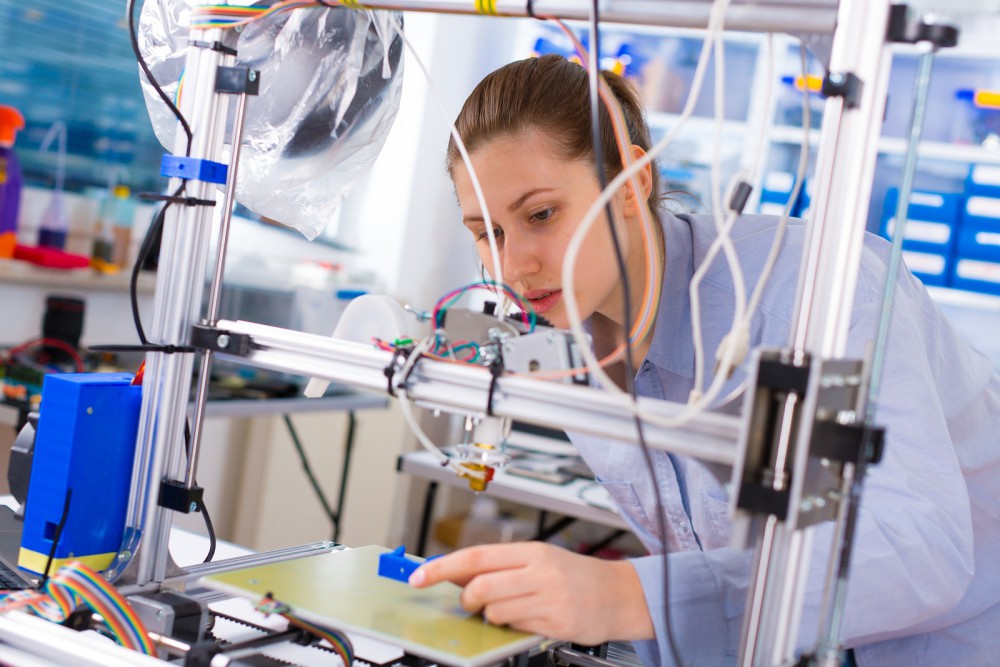
There’s been a leap forward in medicine at Wake Forest University, as researchers at the school’s Institute for Regenerative Medicine have developed a 3D printer capable of creating human-scale tissues that can survive transplants into animals.
Early tests suggest that these tissues are possible candidates for eventual human implantation. Such a breakthrough could potentially be used to help people from all over the world who suffer with infirmities to their internal organs and other sensitive tissues.
The team is led by Anthony Atala, a researcher who has made news already for his efforts in 3D printing components of the human bladder. Thus far, Atala and his team have successfully printed muscle structure, bone material, and tissue for a human ear.
“Our research indicates the feasibility of printing bone, muscle and cartilage for patients,” Atala said. “We will be using similar strategies to print solid organs.”
ITOP
3D printed organs have been in the news for a long time, but Atala and his team have done something unprecedented, and they did it using a printer called the ITOP. The name stands for the ‘integrated tissue organ printer.’
Their paper published in the Nature Biotechnology journal describes the device as a printer that can create human-scale tissue constructs of any shape. It’s effective because it can print cell-laden hydrogels together with biodegradable polymers, which are controlled through a complex clinical imaging data system that controls the printer nozzles and creates tissue in whatever shape is needed.
You Might Also Enjoy: Healthcare Dominates U.S. News & World Report’s Best Jobs Rankings
“To demonstrate construction of a human-sized bone structure,” the paper claimed, “we fabricated a mandible fragment in a size and shape similar to what would be needed for facial reconstruction after traumatic injury. Mandible bone defects have an arbitrary shape. We used data from a CT scan of a human mandible defect … to produce a CAD model of the defect shape, with dimensions of 3.6 cm × 3.0 cm × 1.6 cm.”
In the past, issues with structural integrity and cell oxygenation have kept devices like the ITOP from making much headway in the field of printable, usable tissue. However, advancements by Atala and his team allow the ITOP to print tissues that can be kept alive long enough to be accepted by patients’ bodies.
That’s achieved by printing structures with small channels throughout to facilitate the movement of oxygen and nutrients. This allows a patient’s blood vessels to more easily adopt the implant. This a practice that has been successful when previously tested on rats and mice.
The Future of 3D Tissue Printing
No human tests have been conducted to this point, but the experiments on mice have been encouraging. However, more long term testing is required. Atala and his researchers have only a few months of data on their current test subjects, but once they study the long term progress of the implanted tissues, testing will move forward with clinical-grade human cells with a more diverse variety of cell types.
“When printing human tissues and organs,” Atala said, “we need to make sure the cells survive, and function is the final test.”


